

Understanding a dog’s body is crucial. It unlocks insights into a dog’s health and behavior. This guide simplifies complex dog anatomy. Basic anatomy knowledge helps owners identify potential health issues early. Owners appreciate their dog’s physical capabilities. This deepens the bond with their dog. Become a more informed, attentive pet parent for your dog.
Key Takeaways
Learn about your dog’s body to understand its health and behavior better.
A dog’s head has special senses like super hearing and smell, helping it understand the world.
The torso holds important organs like the heart and lungs, protected by the spine and ribs.
Dog legs are strong for movement, but watch for common injuries like hip problems.
A dog’s tail helps it talk and keep balance, showing its feelings and helping it move.
Understanding Dog Anatomy: An Overview
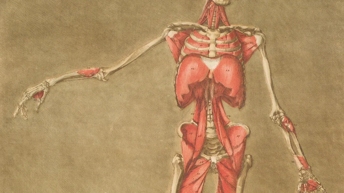
Understanding the fundamental structure of a dog provides valuable insights into its health and capabilities. A dog’s body is a complex and efficient biological machine. Its skeletal system, for instance, forms the core framework. This framework typically contains an average of 319 individual bones. Variations in bone count can occur due to differences in tail length, breed, and the presence of dewclaws. Complementing this bony structure, a dog’s muscular system includes approximately 700 muscles. These muscles enable movement, support, and various bodily functions. This intricate design allows dogs to perform a wide range of activities, from running and jumping to subtle facial expressions.
External Dog Anatomy Regions
External dog anatomy refers to the visible parts of a dog’s body. These regions include the head, neck, torso, limbs, and tail. Each external part plays a specific role in a dog’s interaction with its environment and its overall physical presentation. Observing these external features helps owners identify breed characteristics and potential physical anomalies. Understanding these visible parts of a dog is the first step in appreciating its unique physical form.
Internal Systems Basics
Beyond the visible exterior, a dog’s body houses several vital internal systems. These systems work together to maintain life and function. The seven primary body systems are:
Skeletal System: Provides support, protection, and allows movement.
Muscular System: Enables movement, maintains posture, and produces heat.
Cardiovascular System: Circulates blood, oxygen, and nutrients throughout the body.
Respiratory System: Facilitates breathing and gas exchange.
Digestive System: Processes food, absorbs nutrients, and eliminates waste.
Nervous System: Controls and coordinates all bodily functions, including senses and behavior.
Reproductive System: Responsible for the continuation of the species.
Each system is crucial for the dog’s overall health and well-being. A basic understanding of these internal systems helps owners grasp the complexity of dog anatomy and appreciate how each component contributes to a healthy, active dog.
Canine Head and Sensory Organs
The dog’s head and its sensory organs are crucial parts of its anatomy. These structures allow the dog to interact with its environment. They help the dog understand the world around it.
Skull and Brain Structure
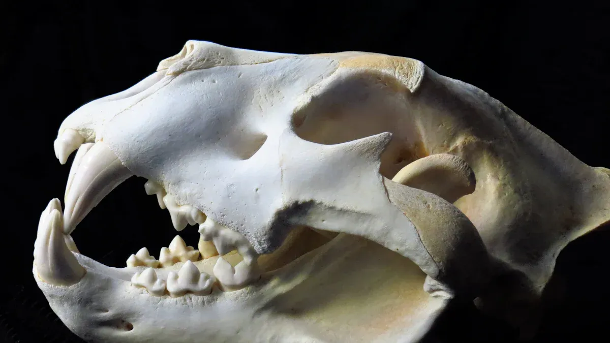
The dog’s head contains its brain. The skull protects this vital organ. The brain is situated within the bony cranium. This is also known as the skull. The neurocranium encloses the brain. The cranium provides protection and support for the brain. It prevents damage during daily activities. This strong bony structure is essential for the dog’s survival.
Eyes and Vision in Dogs
A dog’s eyes provide its vision. Canine senses include sight. Dogs see the world differently than humans. Dogs have dichromatic vision. They primarily see blue and yellow. They struggle to distinguish between red and green. Humans have trichromatic vision. This allows for a broader spectrum of color recognition. Dogs’ visual acuity is not as sharp as humans. They cannot distinguish the same level of detail. Dogs generally have greater peripheral vision than humans. They detect distant movements more easily. Dogs have more rods and fewer cones than humans. This enables them to see much better in dim light or at night. Humans have better color recognition in well-lit conditions. Dogs possess a tapetum lucidum. This reflective layer enhances rod-mediated vision in low light. It reflects unabsorbed light back onto photoreceptors.
Feature | Canine Vision | Human Vision |
|---|---|---|
Color Perception | Dichromatic (blue, yellow) | Trichromatic (red, green, blue) |
Visual Acuity | Lower detail distinction | Higher detail distinction |
Peripheral Vision | Greater | Lesser |
Motion Detection | Easier at distance | Less easy at distance |
Light Sensitivity | Better in dim light/night | Better in well-lit conditions |
Ears and Hearing Capabilities
A dog’s ears are powerful sensory organs. Canine senses include exceptional hearing. Dogs hear a wide range of sounds. Their hearing capabilities surpass human hearing in many ways.
Species | Lower Frequency Limit (Hz) | Upper Frequency Limit (Hz) |
|---|---|---|
Human (young) | 20 | 20,000 |
Human (aging) | 20 | 12,000 |
Dog | 67 | 45,000 |
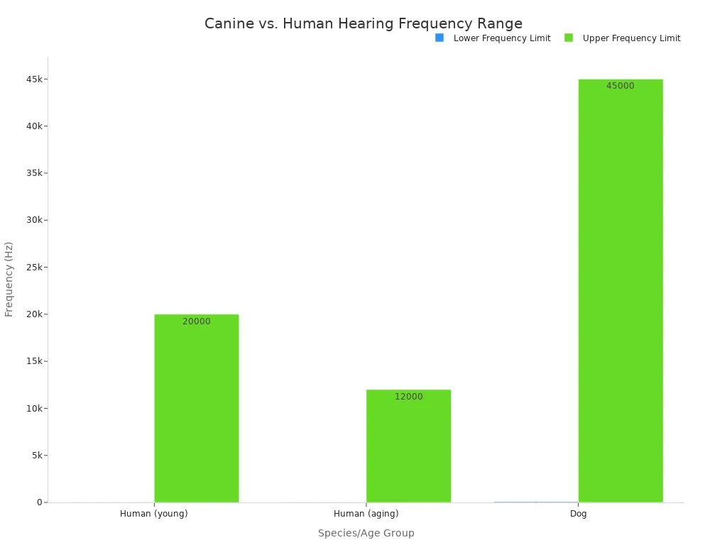
Humans perceive some low-frequency sounds more effectively than dogs. Dogs possess significantly superior hearing sensitivity for high-frequency sounds. This allows them to detect sounds humans cannot hear. The dog’s head moves its ears to pinpoint sound sources.
Nose and Olfactory Power
The dog’s nose is its most remarkable sensory organ. This part of the head is central to its world. Dogs have an incredible sense of smell. Their olfactory power is far superior to humans.
Species | Olfactory Receptors (millions) | Relative to Humans | Area of Receptors |
|---|---|---|---|
Dog | 125 – 300 | ~40 times more | Size of a pocket handkerchief |
Human | ~5 | 1 | Size of a postage stamp |
Humans possess a pair of olfactory receptors. Each is approximately the size of a postage stamp. Dogs’ olfactory receptors can be as large as a handkerchief. The size varies depending on the dog’s breed. This vast number of receptors gives the dog its powerful sense of smell.
Mouth, Teeth, and Tongue
The dog’s mouth contains its teeth and tongue. These are essential for eating and communication. An adult dog has 42 permanent teeth. This is its dental formula: 2x (3/3 I, 1/1 C, 4/4 P, 2/3 M).
Incisors (I): 3/3
Canines (C): 1/1
Premolars (PM): 4/4
Molars (M): 3/2
Total: 42 teeth
The incisors bite and cut food. The canines grasp and tear. Premolars and molars grind food. The tongue helps with eating, drinking, and grooming. The dog’s head uses these structures for many daily functions.
Dog Torso: Core Systems and Structure
The dog’s torso forms the central part of its body. It houses vital organs and provides structural support. This section explores the core systems and structures within a dog’s main body.
Spine and Rib Cage
The spine and rib cage provide crucial support and protection for a dog’s internal organs. The canine spine supports the animal’s weight. It also encases the spinal cord. This provides protection to this vital neurological structure. The spine extends from the base of the neck down through the torso. The rib cage surrounds the chest cavity. The ribs play a crucial role in limiting the movement of the thoracic spine. They also safeguard the internal organs located within the thoracic cavity. Strong muscles connect the neck to the upper rib cage.
Chest Cavity Organs
The chest cavity, or thoracic cavity, contains major organs. These include the heart and lungs. The chest cavity begins below the neck. Dogs possess two large lungs. These lungs divide into lobes. They have a spongy appearance. This is due to a system of delicate bronchioles. These bronchioles end in thin-walled alveoli. The alveoli are the sites of gas exchange. The diaphragm is a muscular structure. It divides the peritoneal cavity from the pleural cavity. It aids the lungs during inhalation. The heart sits within a pericardial cavity. Fluid fills this cavity. The fluid reduces friction. The heart has a right and left side. Each side divides into two chambers: atria and ventricles. The lungs are large, paired organs. They occupy the thoracic cavity. They lie on either side of the mediastinum. Each lung divides into several lobes. The right lung has cranial, caudal, middle, and accessory lobes. The left lung has cranial and caudal lobes. A smooth epithelial layer covers them. The space between the pleura lining the thorax and the pleura lining the lungs is the pleural cavity. This cavity also fills with fluid to reduce friction during breathing. Lungs have a vast surface area for gas exchange. Oxygen diffuses into capillaries. Carbon dioxide diffuses into the air in the alveoli. Oxygen then binds to hemoglobin. It transports to cells. Major blood vessels and nerves also pass through the neck to supply the chest.
Abdominal Cavity Organs
The abdominal cavity follows the chest. It extends from the diaphragm to the pelvic region. This area contains many digestive and excretory organs. These include the stomach, intestines, liver, kidneys, and pancreas. These organs work together to process food, absorb nutrients, and eliminate waste. The abdominal wall protects these organs. It consists of muscles and connective tissue.
Skin and Coat Health
A dog’s skin and coat are its first line of defense. They protect against environmental elements. They also regulate body temperature. A dog’s skin covers its entire body, from the tip of its nose to its tail, including the neck. Maintaining good skin and coat health is vital for a dog’s overall well-being. Several common dermatological conditions can affect a dog’s skin and coat:
Bacterial Dog Skin Infection (Pyoderma): Staphylococcus bacteria often cause this. It arises when the skin barrier is compromised. Symptoms include red bumps, pustules, crusty skin, hair loss, and intense itching.
Fungal Dog Skin Infection (Malassezia Dermatitis): This occurs due to yeast overgrowth. It often happens in warm, moist areas. It leads to greasy, scaly patches with a musty odor and severe itching.
Hot Spots (Acute Moist Dermatitis): These are rapidly developing, moist, inflamed skin areas. Excessive licking, scratching, or chewing causes them. Allergies or insect bites often trigger them. They appear red, warm, and moist with matted fur.
Parasitic Dog Skin Infection (Mange): Microscopic mites cause mange. It results in severe irritation, inflammation, intense itching, hair loss, and scaly patches. Both demodectic and sarcoptic types exist.
Seborrheic Dermatitis: This condition affects the skin’s oil glands. It presents as dry (sicca) or oily (oleosa) scaly patches with an unpleasant odor. Certain breeds are more prone to it.
Allergic Dermatitis: An immune system overreaction to allergens causes this. It leads to intense itching, redness, and inflammation. It can cause secondary infections, scaly patches, hot spots, and chronic ear infections.
Dog Limbs and Movement
A dog’s legs are marvels of biological engineering. They consist of a complex network of muscles, bones, joints, ligaments, tendons, blood vessels, and nerves. This intricate design allows for a wide range of movements, from a gentle stroll to a powerful gallop. The physical construction, or conformation, of a dog significantly influences its movement and athletic capabilities. Understanding the dog leg anatomy helps owners appreciate their dog’s physical prowess and identify potential issues.
Forelegs and Shoulders
A dog’s forelegs and shoulders provide support, balance, and shock absorption. The structure of the front leg anatomy is crucial for a dog’s overall mobility. Unlike humans, a dog does not possess a bony clavicle. Instead, it has a vestigial structure embedded within the shoulder muscles. This structure is not connected to other bones. This anatomical adaptation allows for greater flexibility and range of motion in the forelimbs. It benefits running and leaping by enabling free scapular movement and increasing stride length.
The elbow joint is located below the chest. It is a hinge joint where three bones meet: the humerus (long bone of the upper foreleg), and the radius and ulna (bones of the lower leg). The wrist joint, or carpus, sits between the elbow joint and the paw on the front legs. Here, the bones of the upper leg connect with seven small carpal bones. This forms an intricate system crucial for stopping, standing, and movement.
Several muscles contribute to the strength and movement of the forelegs and shoulders:
Deltoid Muscle: This muscle covers the shoulder joint. It is involved in flexing the limb and lifting the humerus. It originates on the scapular spine and acromion and inserts on the deltoid tuberosity and humeral crest.
Supraspinatus Muscle: This muscle originates on the supraspinous fossa of the scapula spine. It inserts at the greater tubercle of the humerus. It aids in extension and flexion and stabilizes the shoulder joint.
Infraspinatus Muscle: This muscle is found in the infraspinous fossa of the scapula. It functions in flexion and abduction of the forelimb. It originates at the infraspinous fossa and scapula spine and inserts on the humerus. It works with the supraspinatus for shoulder stabilization.
Subscapularis Muscle: This muscle is situated on the subscapular fossa. It helps in internal rotation of the humerus and provides joint stability. It inserts at the coracobrachial muscle on the lesser tubercle of the humerus. It adducts and extends the shoulder and aids in flexion.
Coracobrachialis Muscle: This muscle originates from the distal part of the scapula. It inserts at the proximal part of the humerus. It stabilizes the joint and adducts the shoulder.
Teres Major and Minor Muscles: The Teres Major originates at the caudal margin of the scapula and inserts at the body of the humerus. The Teres Minor originates at the distal infraspinous fossa and infraglenoid tubercle, inserting at the teres minor tuberosity. Both muscles contribute to shoulder stability and flexion.
The canine shoulder joint differs significantly from the human shoulder joint:
Feature | Canine Shoulder Joint | Human Shoulder Joint |
|---|---|---|
Glenoid Labrum (GL) | Not all three zones (transition, shifting, meniscoid fold) present in all joint segments; less important as a passive stabilizer due to height and width. | Consists of three zones (transition, shifting, meniscoid fold); important passive stabilizer. |
Joint Ligaments | Differences exist compared to humans. | Differences exist compared to canines. |
Tendons | Differences exist compared to humans. | Differences exist compared to canines. |
Rotator Cuff | Lacks a rotator cuff. | Possesses a rotator cuff. |
Hindlegs and Hips
A dog’s hindlegs and hips provide the primary power for forward motion. The hind leg anatomy includes several key bones. These bones comprise the hip, knee (stifle), and lower leg. The primary bones comprising a dog’s hindlegs and hips include:
Hip Bones: Ilium, Pubis, Femur, Sacrum bone, Acetabulum, Pubic symphysis.
Knee (Stifle) Bones: Femur, Tibia, Patella (kneecap).
Hip dysplasia is a common orthopedic condition affecting a dog’s hips. It involves abnormal development of the hip joint. This condition can lead to pain, arthritis, and lameness. Canine hip dysplasia shares similarities with late-onset adolescent/adult acetabular dysplasia in humans.
Here are some characteristics of canine hip dysplasia:
Characteristic | Prevalence/Observation |
|---|---|
Overall CHD Prevalence | 15.56% |
Female Predominance | Slightly higher in females (OR 1.05), but with large variations (e.g., 3.4 times more frequent in female Polish Tatra Sheepdogs, 1.6 times more frequent in male Afghan Hounds) |
Laterality | Usually bilateral (67%), unilateral in 33% of cases |
Seasonal Variation (Birth) | More prevalent when born in winter and spring (OR 1.13 for winter, 1.14 for spring) |
Latitudinal Variation | More common in southern latitudes (OR 2.12) |
Breed Variation (Examples) | Ranges from 77.7% in Bulldogs to 0% in Italian Greyhounds (North America); 59.7% in Cane Corso to 3.9% in Siberian Husky (France); 70.6% in Doberman Pinschers to 50% in Golden Retrievers (Turkey) |
AKC Groups | Working dogs had highest risk (OR 1.882) |
FCI Groups | Pinscher/Molossoid group had highest risk (OR 4.168) |
This table shows the prevalence and variations of hip dysplasia across different factors. The condition is often bilateral, meaning it affects both hips. Certain breeds and environmental factors can increase a dog’s risk of developing hip dysplasia.
Detailed Dog Leg Anatomy
The dog leg anatomy is a complex system. It includes bones, muscles, joints, ligaments, and tendons. These components work together to facilitate movement. Ligaments connect bones to other bones. They provide joint stability during activity. Tendons connect muscles to bones. They enable muscular-controlled movements. Both tendons and ligaments are vital supporting elements in a dog’s body structure. They work in an integrated manner with joints, muscles, and bones to facilitate movement. The weakening of any of these tissues, whether due to injury, aging, or chronic inflammation, can lead to a complete breakdown of the dog’s mobility system. This compromises both protection and performance.
Conformation, the physical construction of a dog, significantly impacts its gait and athletic performance.
Overall body size and shape significantly impact performance. Heavier dogs are slower to accelerate, turn, and decelerate. However, they possess more power for pulling or supporting tasks.
Front limb structure, including angulation (shoulder and elbow joints), scapular angle, and humerus length, dictates reach, balance, and stability. Well-angulated limbs allow for longer strides, reducing energy expenditure. They also provide better shock absorption. Straight front legs or upright shoulders (less than 30°) lead to quicker fatigue and increased susceptibility to injuries.
Rear limb structure, particularly angulation and pelvis attachment, provides the primary power for forward motion. The degree of angulation varies by breed. For example, Irish Setters have abundant angulation for covering ground efficiently. Guarding breeds often have more upright limbs for stability. More angulated hind limbs, as seen in some modern German Shepherds, can enable faster movement. This comes at the cost of maneuverability and stability.
Differences in gait patterns observed in agility weave poles correlate with variations in conformation and size among canine athletes. Taller dogs are less likely to use a ‘four feet in, four feet out’ (FFH) technique. This is possibly due to their larger body size and increased stride length. This favors ‘four feet in, two feet out’ (FFSS) or ‘four feet in, two feet in, two feet out’ (FFDS) techniques. Training dogs with shorter natural strides to use techniques not suited to their conformation could negatively impact performance and increase injury risk. Variability in conformation, even within breeds like Border Collies, affects movement. This is especially true in highly physical activities like agility.
Limb straightness, when viewed from the front or rear, and convergence under the body are crucial for biomechanical efficiency. They prevent weight shifting and maximize muscular energy for forward drive. Some breeds sacrifice trotting efficiency for other performance aspects. For instance, ‘cow-hocked’ herding breeds have adducted tarsi and abducted paws. This is an adaptation for improved stability when lying down and standing up. It also reduces rear leg motion during sharp turns required for herding. The arched lumbar spine of a racing Greyhound allows for a much longer stride length at the gallop. It enables the pelvic limbs to reach far forward when the spine is flexed. However, this conformation reduces step length at a trot due to the more vertical slant of the pelvis. This restricts full rearward extension of the pelvic limb unless the spine is fully extended.
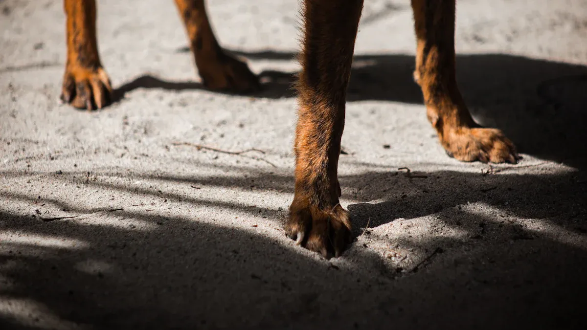
Paws, Claws, and Dewclaws
A dog’s paws are complex structures. They provide traction, absorb shock, and protect the bones and joints. The parts of a dog leg that make up the paw include several types of pads and claws.
Digital pads: These pads are located under each toe. They absorb shock, provide traction, and help distribute load during movement.
Metacarpal/metatarsal pad: This large central pad bears the majority of the body’s weight. It absorbs impact during activities like running or landing.
Carpal pad: This pad is found on the front limbs. It assists in braking, turning, and maintaining control when moving downhill.
Dewclaws: These can help stabilize the limb during agility maneuvers. They may reduce torque on the wrist joint during turns or descents.
Paw pads feature a thick keratinized outer layer. They have an underlying cushion of fat and connective tissue. This structure functions as a shock absorber. It provides insulation from rough or cold surfaces. The pads also contain sweat glands and dense sensory receptors. These support thermoregulation and proprioception (awareness of limb placement and terrain).
Claws are made from keratin. They provide traction across various terrains. They aid in proper posture and assist in digging and tearing. The dewclaw is similar to a human thumb. It is found on the back inner side of the paws, usually the front. While its primary purpose in domestic dogs is largely unknown, it offers additional grip when chewing on objects.
Dewclaws serve several purposes in different dog breeds:
Support and Traction: Dewclaws provide support to the limb when dogs turn. This is especially beneficial for dogs in agility or other sports. They also offer extra traction and help stabilize the carpal (wrist) joint at high speeds. This is particularly useful when turning or on slippery surfaces.
Stabilization: When dogs run at a gallop or canter, dewclaws can touch the ground. This helps stabilize the carpus (wrist) of the lead leg.
Grip and Manipulation: Some dogs use their dewclaws for tasks like climbing, scratching, cleaning their teeth, and gripping bones or chews while eating.
Common Dog Leg Injuries
Dogs can experience various common dog leg injuries. These injuries can affect their mobility and overall well-being. Understanding these issues helps owners seek timely veterinary care. A broken bones in dogs is a serious injury. It requires immediate medical attention. Other common dog leg injuries include sprains, strains, and ligament tears.
Cruciate ligament tears are among the most frequent common dog leg injuries. The recovery process for a cruciate ligament tear is extensive:
First 24-48 Hours: Focus on comfort, calmness, and consistent pain medication. Limit movement to essential bathroom breaks. Monitor for complications.
Weeks 1-2 (Critical Rest Period): Strict rest is vital for initial healing. Confine the dog to a crate or small room. Prevent jumping or running. Use a short leash with a sling for brief bathroom trips. Monitor the surgical site daily.
Weeks 2-8 (Progressive Recovery): Begin controlled daily activities. Introduce low-impact exercises and gradually longer leashed walks as guided by the vet. Professional physical therapy may start during this period.
Months 2-3 (Building Strength): Continue strengthening the leg with increasingly challenging but controlled activities. Longer walks and more advanced physical therapy exercises may be introduced. Still avoid sudden or strenuous movements.
3-6 Months (Return to Normal Activity): Gradually reintroduce regular activities under supervision. Implement ongoing exercise and weight management strategies to protect the repaired joint. Consider preventive care.
This detailed recovery timeline highlights the importance of patience and consistent care for a dog recovering from a significant leg injury.
Tail and Rear Anatomy
Tail Structure and Function
A dog’s tail is more than just an extension of its body. It serves important roles in communication and balance. The tail typically contains between six and 23 highly mobile vertebrae. Versatile muscles surround these vertebrae. These muscles enable fine movements of the tail. They allow lifting, side-to-side motion, and drawing the tail down. Caudal muscles originate from the lumbar vertebrae and sacrum. They attach exclusively to the tail vertebrae. Tendons connect these muscles to the tail vertebrae. The most posterior tendons attach to the last tail vertebrae. Four to seven paired nerves innervate the tail muscles. Some musculature also comes from muscles linked to the rectum, anus, and pelvic diaphragm.
The canine tail is a visible communication tool. Its speed, position, and carriage convey emotions. A worried dog carries its tail lower. A fearful dog tucks it. An alert, confident dog holds it higher and still. The tail works with facial expressions and body movement for communication. Dogs also use their tails as a counterbalance. This helps them during running, leaping, and turning. Strong muscles in the hips, pelvis, and tail move the tail as needed. This maintains balance during movement, such as when jumping or landing. In swimming, a dog’s tail acts as a rudder for steering. It moves from behind to one side during turns. It also helps maintain balance against forces like waves.
Rear End and Pelvic Region
The rear end and pelvic region form a crucial part of a dog’s structure. This area provides power for movement. The os coxae, or hip bone, is a key bone in this region. Other important bones include the pelvis, femur, sacrum bone, acetabulum, and pubic symphysis. Many muscles support the pelvic region. Thigh muscles include the biceps femoris, semitendinosus, semimembranosus, sartorius, gracilis, pectineus, and adductor muscles. Rump muscles include the tensor fasciae latae, superficial gluteal, middle gluteal, piriformis, deep gluteal, and articularis coxae muscles.
Several health issues can affect a dog’s pelvic region. Hip dysplasia is a primary arthropathy of the pelvic limb. Hip luxation also affects the hip. Other conditions include cranial cruciate disease, meniscal injury, and patellar luxation. Iliopsoas strains are muscle disorders. These often occur during eccentric contraction. Gracilis and semitendinosus/semimembranosus myopathy are also muscle disorders. Pelvic bladder is another condition. It involves displacement of the bladder. This can affect its size or the position of the urethra. This is a congenital condition. It is more common in intact female dogs.
Understanding dog anatomy is vital for a dog’s overall well-being. This guide provided insights into a dog’s structure, from its skeletal system to its sensory organs. This knowledge of dog anatomy empowers owners to be more observant and proactive regarding their dog’s health. Owners can better appreciate their dog’s physical capabilities. They can also identify potential issues early. Continue learning about your dog. Always consult a veterinarian for any health concerns. A deeper connection with your canine companion comes from understanding its unique structure.
FAQ
What is the primary function of a dog’s dewclaws?
Dewclaws help dogs grip objects. They provide extra traction when running or turning. Some dogs use them to stabilize their wrist joint during high-speed maneuvers. They can also assist in climbing or holding items.
How does a dog’s vision compare to human vision?
Dogs see fewer colors than humans. They primarily see blue and yellow hues. Dogs have better night vision due to more rods in their eyes. They also detect movement more easily. Humans see more detail and a wider color spectrum.
What are the main functions of a dog’s tail?
A dog’s tail serves two main purposes. It acts as a communication tool, conveying emotions through its position and movement. The tail also helps with balance. It acts as a counterbalance during running, jumping, and turning.
What is hip dysplasia in dogs?
Hip dysplasia is a condition where the hip joint develops abnormally. The ball and socket do not fit together correctly. This causes pain, arthritis, and lameness. It is a common orthopedic issue, especially in larger breeds. 🦴